Permitting the passage of x rays – Permitting the passage of X-rays, a topic of immense scientific significance, embarks us on a journey of discovery, shedding light on the principles, applications, and implications of this remarkable technology.
X-rays, with their ability to penetrate matter and reveal hidden structures, have revolutionized various fields, including medicine, security, and industrial inspection. This article delves into the fascinating world of X-rays, exploring their types, imaging techniques, and diverse applications, while also addressing potential risks and alternative imaging modalities.
Types of X-rays

X-rays are a form of electromagnetic radiation, similar to visible light but with much shorter wavelengths. They are used to create images of the inside of the body, and are particularly useful for diagnosing and treating bone and joint problems.
There are different types of X-rays, each with its own advantages and disadvantages.
Conventional X-rays
- Produce a two-dimensional image of the body part being examined.
- Used to diagnose a wide range of conditions, including bone fractures, joint problems, and chest infections.
- Relatively inexpensive and widely available.
- Can expose the patient to a relatively high dose of radiation.
Computed tomography (CT) scans
- Produce cross-sectional images of the body, which can be used to create three-dimensional reconstructions.
- Used to diagnose a wide range of conditions, including cancer, heart disease, and stroke.
- More expensive and less widely available than conventional X-rays.
- Can expose the patient to a higher dose of radiation than conventional X-rays.
Fluoroscopy
- Produces a real-time image of the body, which can be used to guide procedures such as surgery and angiography.
- Used to diagnose a wide range of conditions, including gastrointestinal problems, heart disease, and vascular disease.
- Can expose the patient to a relatively high dose of radiation.
Mammography, Permitting the passage of x rays
- A type of X-ray used to screen for breast cancer.
- Uses a low dose of radiation to produce images of the breast tissue.
- Can be used to detect breast cancer at an early stage, when it is most treatable.
Equipment Used in X-ray Imaging: Permitting The Passage Of X Rays

X-ray imaging requires the use of specialized equipment, including:
X-ray tube
- Generates X-rays by passing an electrical current through a vacuum tube.
- The voltage applied to the tube determines the energy of the X-rays produced.
X-ray detector
- Converts X-rays into an electrical signal.
- The type of detector used depends on the type of X-ray imaging being performed.
Computer
- Processes the electrical signal from the detector to create an image.
- The computer can also be used to manipulate the image, such as by adjusting the contrast or brightness.
Patient positioning equipment
- Used to position the patient correctly for the X-ray.
- This equipment includes tables, chairs, and other devices.
Procedure for X-ray Imaging

The procedure for X-ray imaging is relatively simple and painless.
- The patient will be asked to remove any metal objects from their body, such as jewelry or clothing with metal buttons.
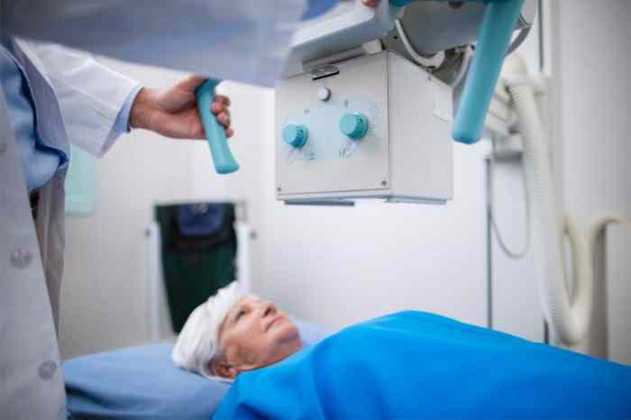
- The patient will be positioned on the X-ray table.
- The X-ray tube will be positioned over the area of the body being examined.
- The X-ray tube will be activated, and the X-rays will pass through the body and onto the detector.
- The detector will convert the X-rays into an electrical signal, which will be processed by the computer to create an image.
The entire procedure usually takes only a few minutes.
Interpretation of X-ray Images
X-ray images can be interpreted by a radiologist, a doctor who is specially trained in reading and interpreting medical images.
The radiologist will look for abnormalities in the X-ray image, such as:
- Fractures
- Dislocations
- Tumors
- Infections
The radiologist will then write a report that describes the findings of the X-ray.
Detailed FAQs
What are the different types of X-rays?
X-rays encompass a wide range, including conventional radiography, fluoroscopy, computed tomography (CT), and digital radiography. Each type offers unique advantages and applications.
How do X-rays contribute to medical diagnosis?
X-rays provide valuable insights into the internal structures of the body, aiding in the diagnosis of fractures, tumors, infections, and other medical conditions.
Are X-rays harmful to health?
While X-rays involve exposure to ionizing radiation, the risks are generally low when performed judiciously. However, excessive exposure can increase the risk of radiation-induced health effects.